


 Tribune Group GmbH Inc.
Nationally Approved PACE Program
Provider for FAGD/MAGD credit.
Approval does not imply acceptance by
any regulatory authority or AGD endorsement.
7/1/2024 - 6/30/2028.
Provider ID# 355051
Use of Bio-C Sealer bioceramic cement in sealing accidental perforations
Sponsor: Angelus
Author: Fabio Takemi Ferreira Kamikabeya
Co-authors: Clauber Romagnoli; Douglas Giordani Negreiros Cortez; Renato Interliche.
RESUME
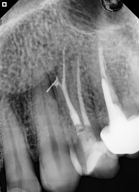
Patient J.E.S., 41 years old, male, referred by an endodontist for evaluation and treatment of palatal root perforation of tooth 24, with file fragment beyond the perforation and periapical lesion.
After isolating the tooth, it was thoroughly irrigated with 2.5% sodium hypochlorite and, with the aid of an apex locator, a measurement of 18 mm was obtained, which was possibly the location of the perforation. Next, a manual file, previously curved to locate the correct path of the canal to the radiographic apex, was used.
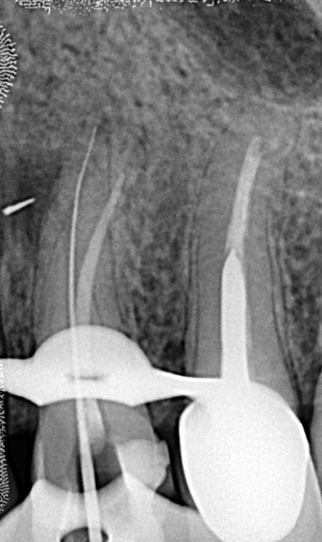
With each new exploration, the 2.5% sodium hypochlorite solution was replaced by abundant irrigation. Once the correct path was located, the apex locator was used again, indicating a working length of 20 mm, which was confirmed after radiography.
The biomechanical preparation of the palatine canal was performed using NiTi rotary files, and after completion of the instrumentation, a final irrigation protocol was applied using endodontic ultrasound with the PUI (passive ultrasonic irrigation) technique.
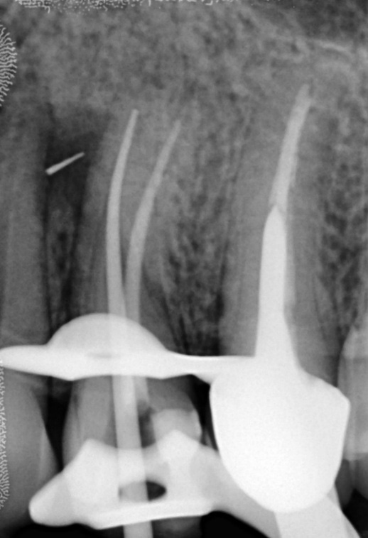
For obturation, the canal was slightly dried with suction tips and obturation was performed using the single cone technique with hydraulic compression. For this purpose, Bio C Sealer bioceramic cement (Angelus, Londrina, Brazil) was used according to the manufacturer’s instructions. The cement was inserted directly from the syringe using the cement’s own applicator tips.
After the filling was completed, the pulp chamber was sealed and a final radiograph was taken, and the patient was instructed to return for follow-up.
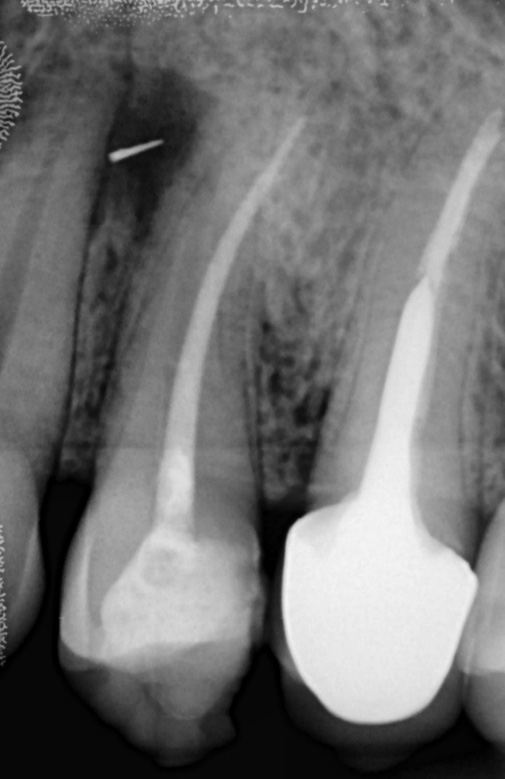
Initial radiograph. Fractured file outside the perforation and presence of periapical lesion.
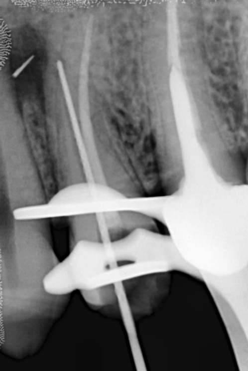
Manual file until perforation.
Recovery of the correct path of the palatine canal with a #10 hand file.
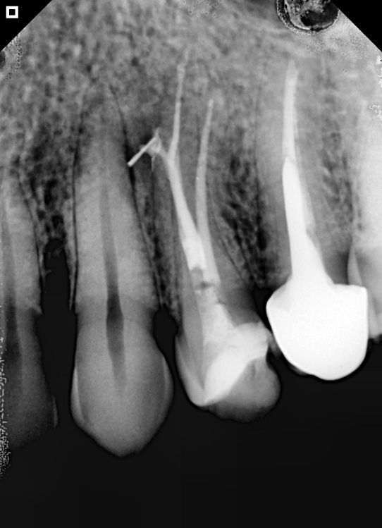
Calibrated gutta-percha cone test.
Finished canal. We see the overflow of bioceramic cement in the perforation.
Radiographic control at 2 months.
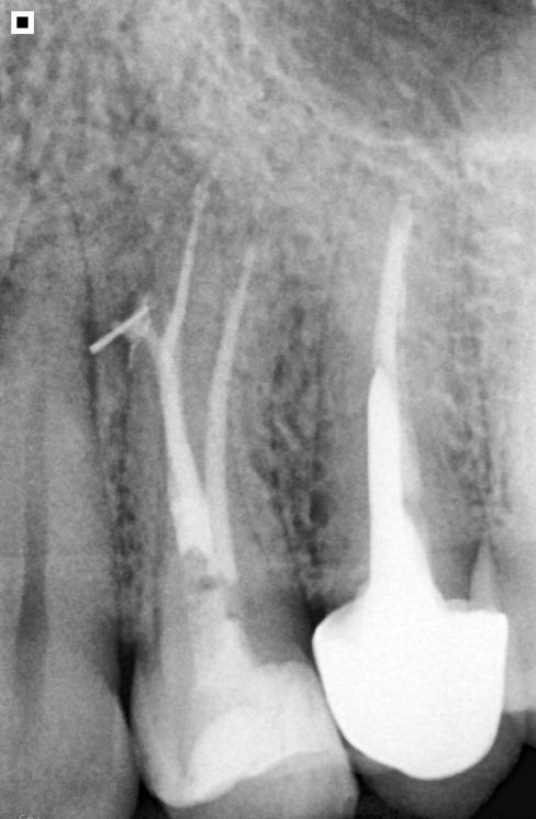
Radiographic control at 5 months.