


 Tribune Group GmbH Inc.
Nationally Approved PACE Program
Provider for FAGD/MAGD credit.
Approval does not imply acceptance by
any regulatory authority or AGD endorsement.
7/1/2024 - 6/30/2028.
Provider ID# 355051
Endodontic Retreatment of Mandibular Molar 46 Using a Finisher File, Bio-C Temp and Bio-C Sealer
Author: Kleber Carvalho
Sponsor: FKG / Angelus
Resume
A 77-year-old male patient was referred for retreatment of tooth 46, which had a poorly conducted endodontic treatment, extensive periapical periodontitis, and an active fistula. The tooth had a prosthetic crown and an extensive fiber post in the distal canal (Figure 01).The fiber post was removed without structural damage to the distal canal, but unfortunately, the small remaining gutta-percha from the apical third was displaced to the periapex (Figure 02).Since the apical foramen was enlarged by the previous treatment, A file Finisher 1 mm was used beyond the apical limit and the extruded gutta-percha fragment was removed (Figure 03).
After the cleaning and re-preparation of the canals, all canals were filled with Bio-C Temp Figure (04).The next session was scheduled for 20 days later. On return, there were no symptoms or restrictions for the obturation phase. The cement used was Bio-C Sealer (Figure 05).
In Figure 06 the comparison between the initial radiograph (March/2025) and a first control, seven months after the end of treatment. The result is very encouraging.
Images
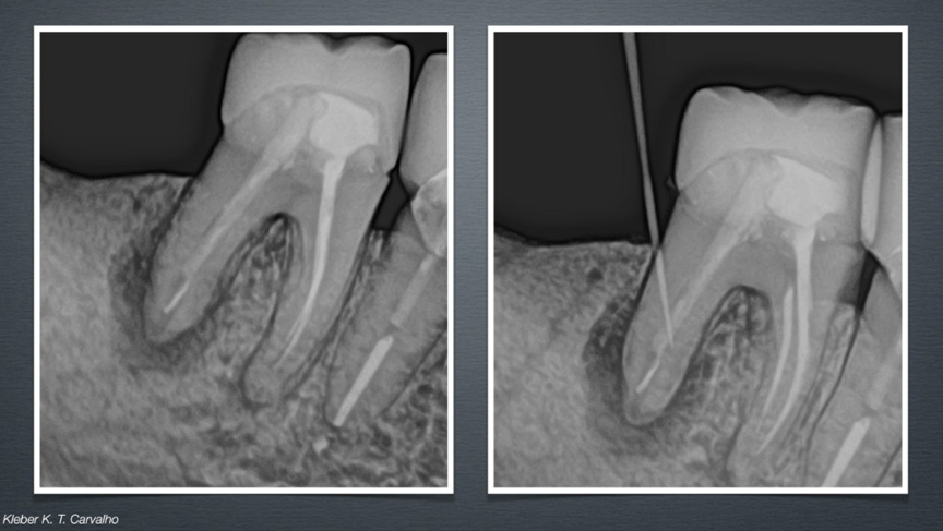
Image 1. Initial X-ray of tooth 46 showing apical periodontitis and an active sinus tract associated with a previous root canal treatment and a fiber post in the distal canal
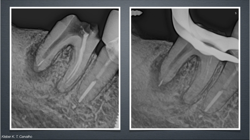
Image 2. Radiograph after fiber post removal, revealing a small gutta-percha fragment displaced into the periapical area
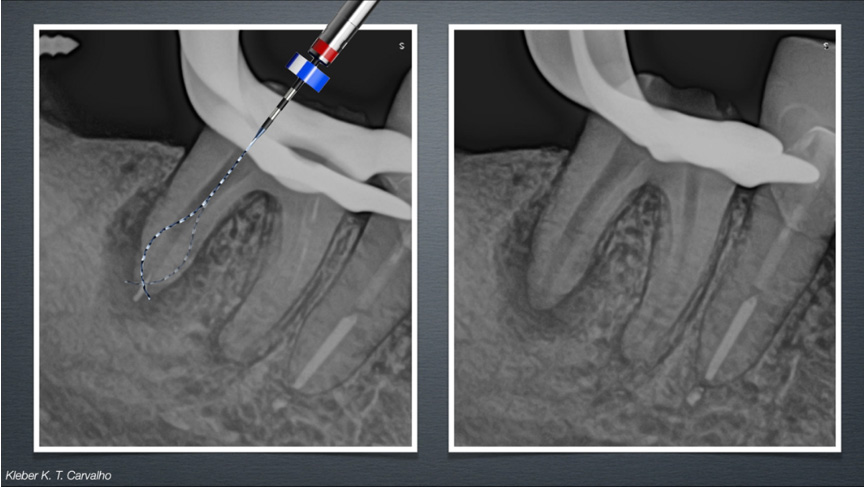
Image 3. Removal of the extruded gutta-percha fragment using a finisher file beyond the apical foramen
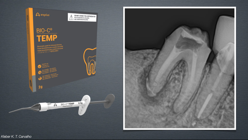
Image 4. Root canals filled with Bio-C Temp used as an intracanal medicament
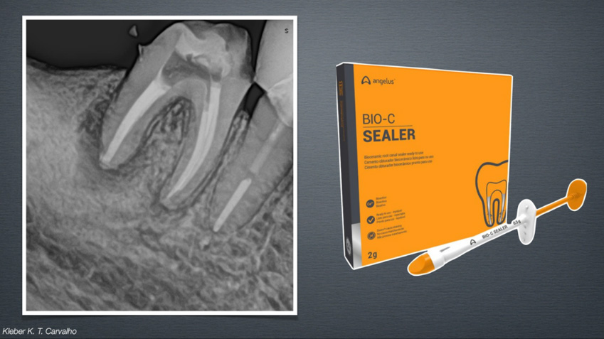
Image 5. Final obturation of all root canals with Bio-C Sealer and gutta-percha cones
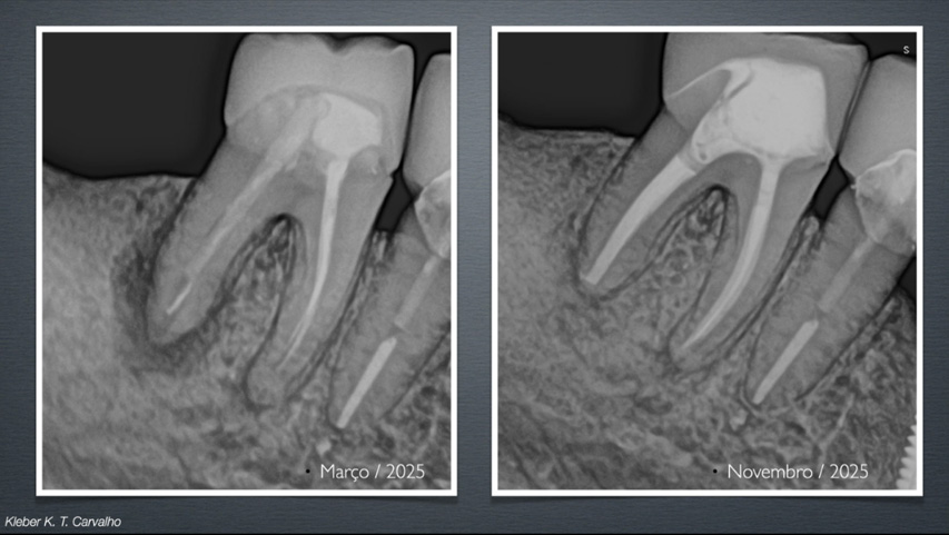
Image 6 – Comparison between the initial radiograph and the 7-month follow-up X-ray, showing evident reduction of the apical radiolucency