


 Tribune Group GmbH Inc.
Nationally Approved PACE Program
Provider for FAGD/MAGD credit.
Approval does not imply acceptance by
any regulatory authority or AGD endorsement.
7/1/2024 - 6/30/2028.
Provider ID# 355051
Does Endodontic Treatment with Bioceramic Cements Accelerate the Recovery of Periapical Lesions? A Case Report
Sponsor: Angelus
Author: Lucas Henrique Gomedi
Co-author: Clauber Romagnoli; Douglas Giordani Negreiros Cortez; Renato Interliche
RESUME
A 25-year-old female patient was referred to the Endodontics Specialization course at the Escola Educação Inteligente – Uningá (Londrina, Paraná, Brazil) for evaluation and endodontic treatment of tooth 46 (lower right first molar). Clinical and radiographic examinations (periapical radiography) were performed, as well as anamnesis, and a coronary opening was found. The tooth was asymptomatic at the time, and the periapical lesion on element 46 was clearly visible with radiography (Figure 1), indicating pulp necrosis.
After evaluation, the access cavity was refined with ultrasonic insert. A rubber dam and a light-curing gingival barrier (Angelus) were used to establish absolute isolation, followed by canal exploration with K#10 hand files and continuous irrigation with a 2.5% sodium hypochlorite solution.
Electronic odontometry was carried out using an Airpex foraminal locator (Angelus). The working length was established at the foramen (zero of the electronic apex locator). Chemical-mechanical preparation was performed using rotary mechanized files. The gutta-percha cone was adjusted to the properly prepared canal (Figure 2). After conometry, each root canal received three applications of 1.7% trisodium EDTA and 2.5% sodium hypochlorite, which were potentiated for 30 seconds at 10% power using ultrasonic insert.
The filling was carried out with Bio-C Sealer bioceramic endodontic cement (Angelus) (Figure 3A). The cement was inserted into the canals using the product’s supplied tips, 4 mm below the working length, and dispensed slightly up to the cervical third. After inserting the cement, gutta-percha cones were inserted and filled using the hydraulic compression technique (Figure 3B).
Following the filling of the canals, the pulp chamber was sealed and filled with calcium phosphate and glass ionomer cement. A follow-up appointment was scheduled after 30 and 90 days. (Figure 4A and 4B).
IMAGES
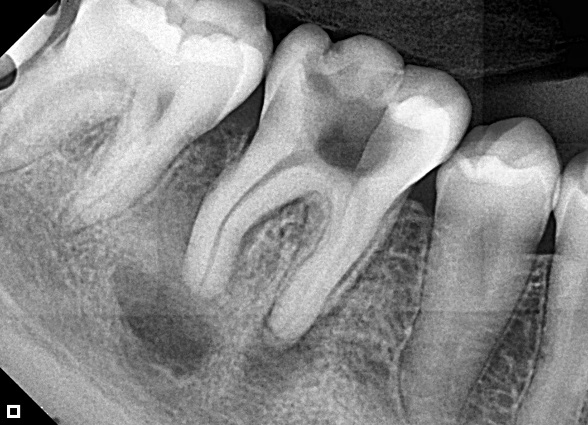
Figure 1: Initial situation
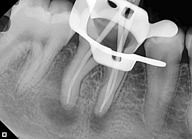
Figure 2: Conometry
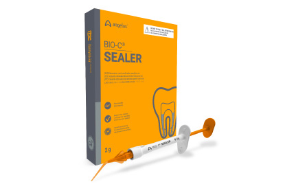
Figure 3: Obturation Cement (A)
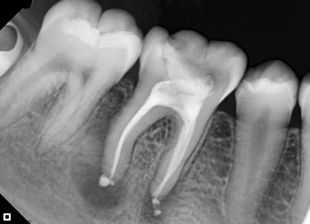
Figure 3: Final radiograph (B)
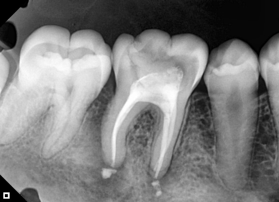
Figure 4: Follow-up with 30 days (A)
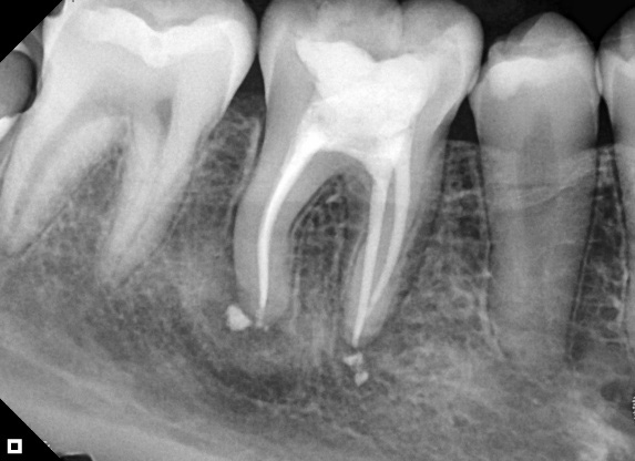
Figure 4: Follow-up with 90 days (B)