


 Tribune Group GmbH Inc.
Nationally Approved PACE Program
Provider for FAGD/MAGD credit.
Approval does not imply acceptance by
any regulatory authority or AGD endorsement.
7/1/2024 - 6/30/2028.
Provider ID# 355051
Cervical Radicular Perforation: Case Report
Sponsor: Angelus
Author: Larissa de Andrade
Co-author: Clauber Romagnoli; Douglas Giordani Negreiros Cortez; Renato Interliche
RESUME
Patient I.N.A.A., 29 years old, female, came to the Escola De Ensino Educação Inteligente (EI Uningá – Londrina, Paraná) after being treated at a private clinic. The patient reported that a routine radiographic examination was performed, and the dentist identified a periapical lesion in tooth 11, in addition to darkening of its color. Endodontic treatment of the tooth was then proposed. At the same location, an attempt was made to access the pulp, but without success. She was referred for a CT scan (Figures 1A and 1B).
At the school clinic, access to the tooth was tried again, but due to the anatomical conditions of this tooth, perforation occurred (Figure 2). After controlling the bleeding in the region, the canal was accessed. Calcium hydroxide was placed as a temporary dressing.
In the second session, absolute isolation was achieved using a rubber dam. The area was irrigated with 2.5% sodium hypochlorite. The canal was located, instrumented, and filled with gutta-percha and Bio-C Sealer bioceramic cement (Angelus) (Figure 3A). The treatment was performed with the aid of an operating microscope to obtain magnification (Figure 3B).
After filling the root canal up to the perforation limit, a hemostatic sponge was placed in the perforation, and Bio-C Repair (Angelus) (Figure 4A) was inserted. A moistened sterile cotton ball and condenser were used to accommodate the cement inside the perforation with minimal pressure to prevent spillage. After sealing, a temporary restoration was performed with glass ionomer cement (Figure 4B). Following this, a follow-up appointment was scheduled for one month later (Figure 5).
IMAGES
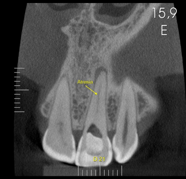
Figure 1: Tomographic image, coronal section (A)

Figure 1: Tomographic image, sagittal section (B)
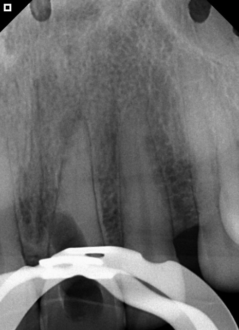
Figure 2: Perforation
Figure 3: Cement sealer (A)
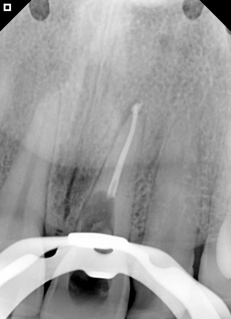
Figure 3: Radiograph after completion of root canal filling (B)
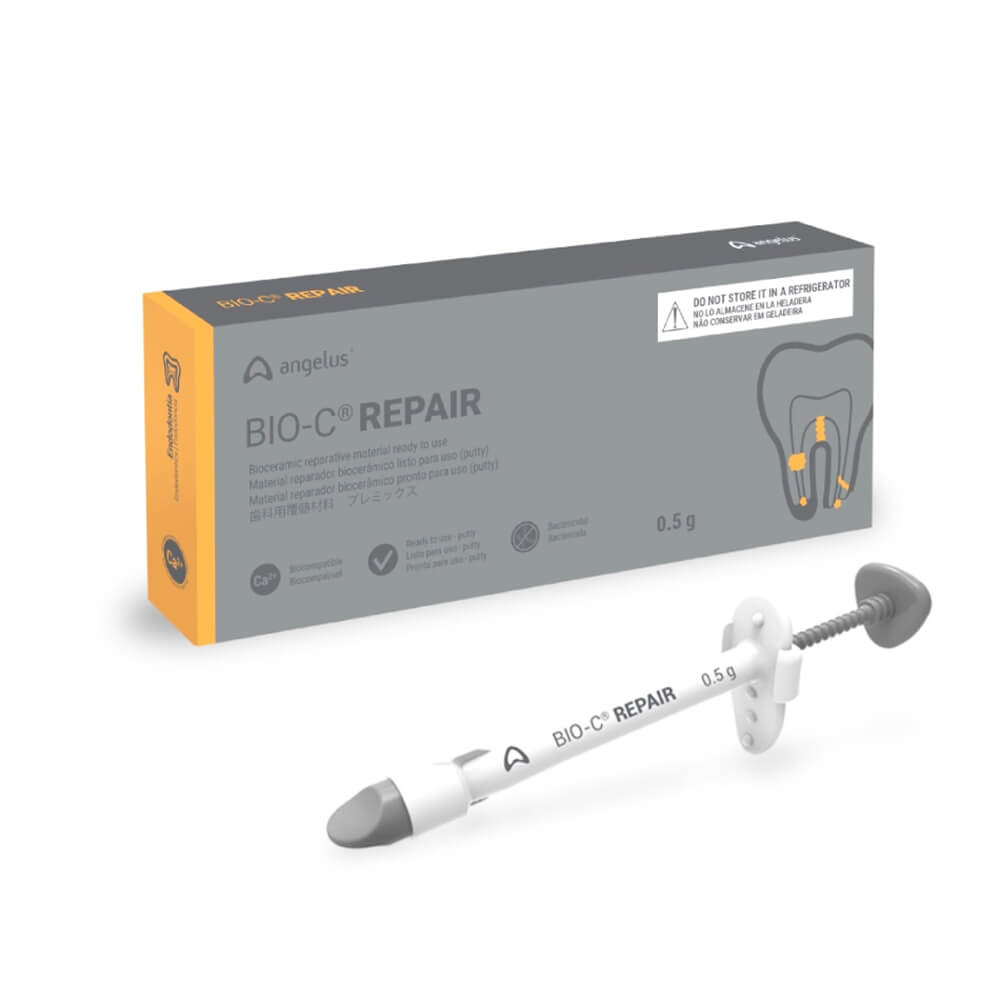
Figure 4: Repair cement (A)

Figure 4: Final radiograph after sealing the perforation (B).
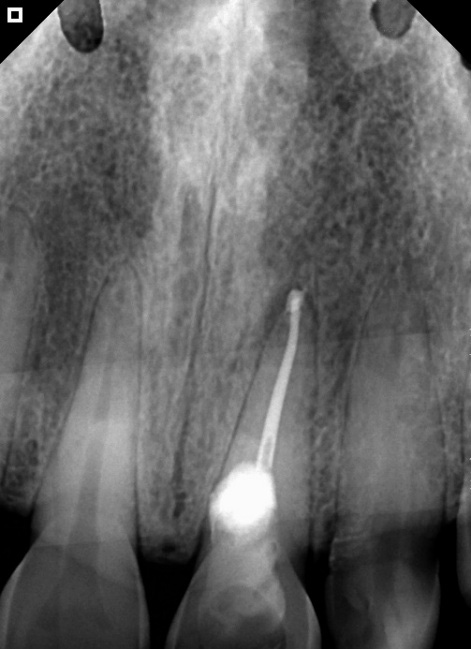
Figure 5: Follow-up radiograph after 1 month