


 Tribune Group GmbH Inc.
Nationally Approved PACE Program
Provider for FAGD/MAGD credit.
Approval does not imply acceptance by
any regulatory authority or AGD endorsement.
7/1/2024 - 6/30/2028.
Provider ID# 355051
BIO-C® TEMP: A new alternative for intracanal medication
Sponsor: Angelus
Author: Clauber Romagnoli
Co-author: Douglas Giordani Negreiros Cortez; Renato Interliche
RESUME
M. M. D., a 34-year-old female patient, visited the dental clinic at Escola Educação Inteligente of Londrina for an evaluation and treatment of tooth 32. During anamnesis and clinical examination, the patient reported that the tooth had an acute abscess approximately a month before (Figure 1).
The first session involved anesthesia, endodontic access correction, absolute isolation, and root canal preparation. During the mechanical chemical preparation, constant irrigation and patency of the canal were used at each file change. The first session concluded with the canal being filled with the intracanal dressing BIO-C® TEMP® (Angelus), followed by a radiograph to confirm that the canal had been filled (Figure 2).
After 30 days, the patient returned and a new periapical radiograph was taken before the intracanal dressing was removed. This radiograph revealed that the periapical lesion had recovered (Figure 3). The intracanal dressing was removed with NaOCl irrigation, followed by a final rinse with EDTA and NaOCl, which was boosted with Easyclean.
As with the first session, any remaining sodium hypochlorite was irrigated with saline solution. The canal was dried using absorbent paper cones and aspirator tips. The gutta-percha cone was selected (Figure 4) and obturation was realized with Bio-C Sealer (Angelus) (Figure 5A and 5B). A follow-up with 90 days was done and showed healing at the apical area (Figure 6A and 6B).
IMAGES
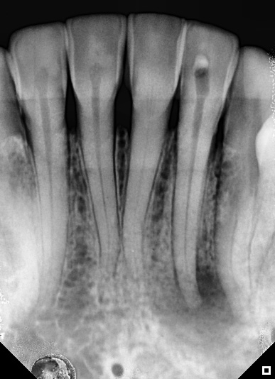
Figure 1: Initial radiograph
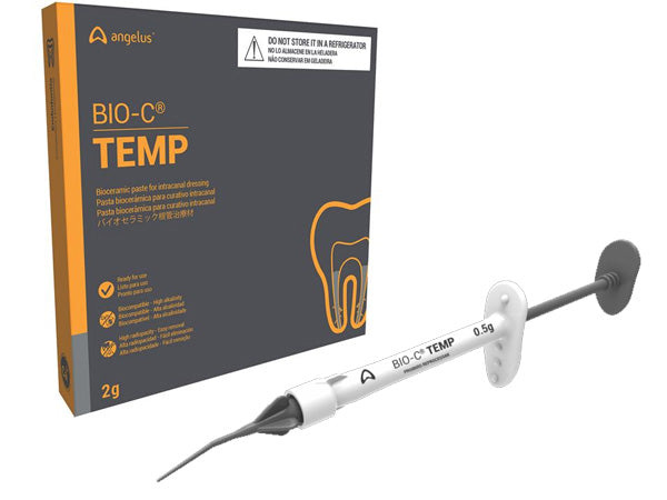
Figure 2: BIO-C® Temp medication applied to the canal after the preparation had been completely carried out
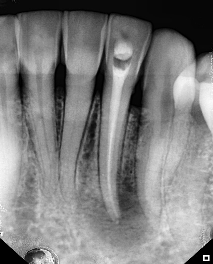
Figure 2: BIO-C® Temp medication applied to the canal after the preparation had been completely carried out
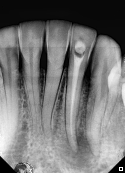
Figure 3: radiographic situation after 30 days. Note the visible regression of the lesion

Figure 4: Guta-percha cone test
Figure 5: Obturation Cement (A)
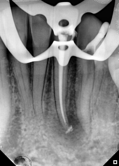
Figure 5: Radiograph to check the quality of the filling (B)
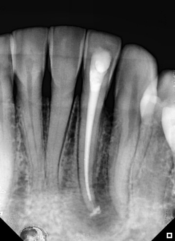
Figure 6: Final radiograph (A)
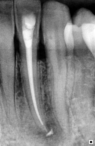
Figure 6: Follow-up after 90 days (B)