


 Tribune Group GmbH Inc.
Nationally Approved PACE Program
Provider for FAGD/MAGD credit.
Approval does not imply acceptance by
any regulatory authority or AGD endorsement.
7/1/2024 - 6/30/2028.
Provider ID# 355051
Apical abces in one visit treatment
Sponsor: EdgeEndo
Author: Prof. Geert Hommez
RESUME
Patient was referred with a buccal swelling in the 2nd quadrant. Crowns on all teeth were placed. Although the crown on 26 is too large at the margins, there is no leakage. The patient does not want me to remove the crown.
Pre-operatieve radiograph show an apical radiolucency on the mesiobuccal root and a small one on the discobuccal root. Diagnosis was apical abces due to pulpal necrosis.
A root canal treatment in one visit was performed through te crown. Four canals were treated. All canals were instrumented with Edge-One-R files (R25 for the buccal canals, R50 for the palatal canal).
The canals were rinsed with 5% NaOCl during and after shaping, followed by EDTA irrigation. Both irrigants were activated ultrasonically, followed by laser cleaning with diode laser.
The canals were dried with sterile paper points. Bioceramic root canal sealer (Edge Sealer) and gutta percha were used with warm vertical condensation.
The acces cavity was closed with fiber-reinforced flowable composite in layers of 1-2mm, followed by a final layer of posterior composite.
IMAGES
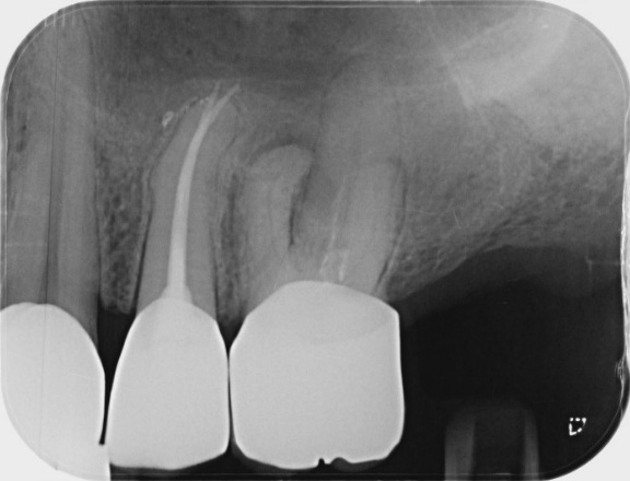
Initial XR
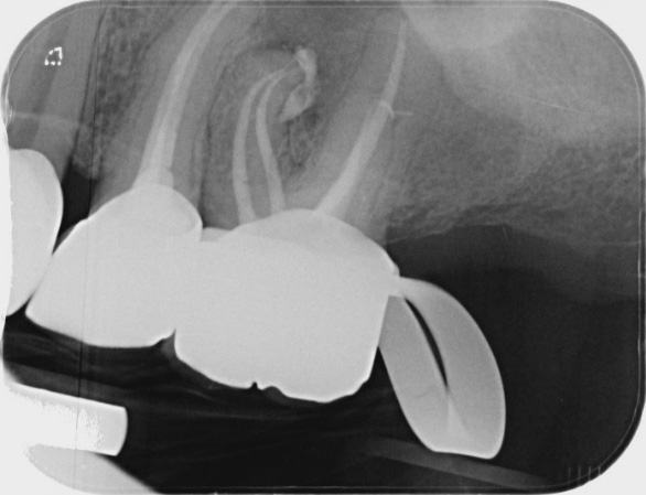
Final XR